Idiopathic Scoliosis (IS) is a complex three-dimensional spasm of the spine, characterized primarily by the curvature of the spine and the rotation of the vertebrae, which not only affects the physique and mobility of the patient, but can also cause back pain, decreased activity, and respiratory impairment. Clinically, the assessment of IS patients is still largely dependent on static imaging methods such as X-rays, CT, MRI, and clinical experience of doctors. Although these methods can reflect well the static shape and curvature of the spine, it is difficult to capture the dynamic and muscular coordination of the spine during actual exercise, so a definitive assessment of recovery quality, dynamic balance, and functional ability is still lacking.
To address these issues, a group of professors from the Beijing University of Industry, Prof. Li Quang, Prof. Dr. Dong Jie, Prof. Dr. Chen, Prof. Dr. Chen, Prof. Dr. Chen, Prof. Dr. Chen, Prof. Dr. Chen, Prof. Dr. Chen, Prof. Dr. Chen, Prof. Dr. Chen, Prof. Dr. Chen, Prof. Dr. Chen, Prof. Dr. Chen, Prof. Dr. Chen, Prof. Dr. Chen, Prof. Dr. Chen, Prof. Dr. Chen, Prof. Dr. Chen, Prof. Dr. Chen, Prof. Dr. Chen, Prof. Dr. Chen, Prof. Dr. Chen, Prof. Dr. Chen, Prof. Dr. Chen, Prof. Dr. Chen, Prof. Dr. Chen, Prof. Dr. Chen, Prof. Dr. Chen, Prof. Dr. Chen, Prof. Dr. Chen, Prof. Dr. Chen, Prof. Dr. Chen, Prof. Dr. Chen, Prof. Dr. Chen, Prof. Dr. Chen, Prof. Dr. Chen, Prof. Dr. Chen, Prof. Dr. Chen, Prof. Dr. Chen, Prof. Dr. Chen, Prof. F. F. F. F. F. F. F. F., and Prof. F. F. F. F. F. F. F. and Prof. and Prof. F. and Prof. and Prof. and Prof. and Prof. and Prof. fellows. had proposed a paper on a focus on a focus on the study on the study on the study on the use of the use of multi-disciplinary and the use of biomedical methods of biomedical sciences in biomedical sciences in biomedical sciences on biomedical sciences, and the study on biomedical sciences on biomedical sciences on biomedical sciences on biomedical sciences, and on biomedical sciences on biomedical sciences, and on biomedical sciences on biomedical sciences, and on biomedical sciences, and related on biomedical sciences, and on biomedical sciences on biomedical sciences, and on the study on the study on health sciences, and on health sciences.
The study as a whole constructed a multi-modal dynamic assessment system aimed at the IS recovery movement, consisting mainly of the semig evaluation modules, SPI evaluation modules, and three parts of the evaluation results and guidance modules. The system first used surface myoelectromagnetic signals to analyze the level of muscle activation of the patient in the recovery operation, the position of the main force muscles and the state of muscle fatigue; then used the motion capture system to obtain information about the spatial trajectory of the movement of the patient, and evaluate from the range of motion and symmetrical stability of the movement; finally, combining the two types of information to establish a rehabilitation confidence coefficient, to quantify whether the patient's movements are regulated and to provide feedback and guidance on existing problems.
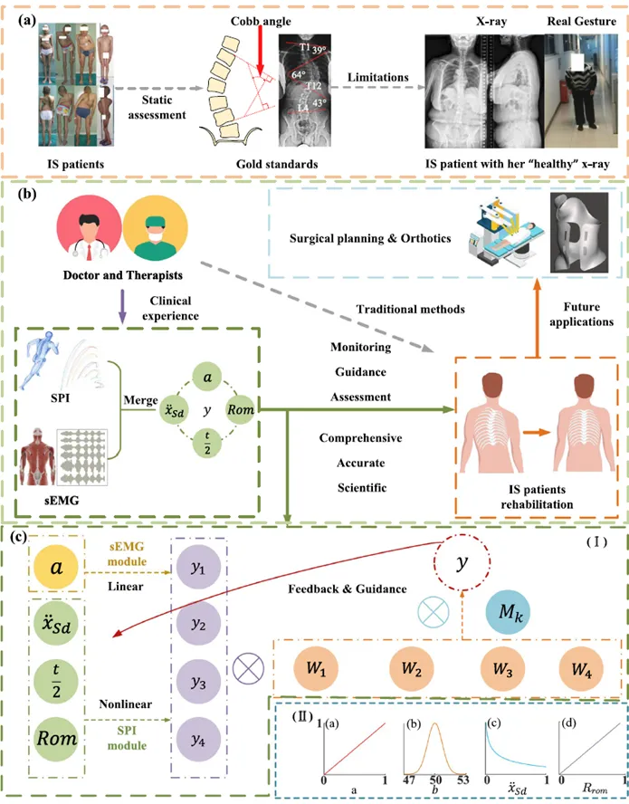
Figure 1 The overall research framework
01.
Multimodal dynamic assessment system
The multi-modal dynamic assessment system, as shown in Figure 2, combines sEMG with SPI to address the problem of making a comprehensive assessment of actions that rely solely on a single piece of information in the recovery process of IS patients.
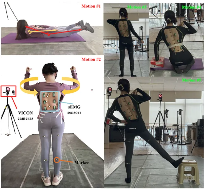
Figure 2 Multimodal dynamic assessment system
The sEMG is primarily used to describe the state of muscle activity in patients performing rehabilitation movements. The researchers placed a total of six wireless electron sensors on each side of the T7, T12 and L3 positions of the subjects' backs to collect spinal muscles at different locations. By comparing the degree of activation of different muscle pathways, it is possible to determine which part of the muscle the patient mainly uses to complete the movement and further determine whether the main force position is consistent with the target training muscle.
SPI is primarily used to describe the state of spatial motion in patients during rehabilitation movements. The study uses the VICON optical motion capture system to obtain three-dimensional spatial information about subjects' body markers and to adapt Gauss functions to the trajectory of rehabilitation movements.
02.
Personalized Spinal Movement Capture Models
The Plug-in Gait model commonly used in the traditional VICON system is difficult to accurately describe the spine shape of an IS patient with distinct lateral curvature and rotation characteristics. Therefore, this study focuses on the spinal curvature characteristics of an IS patient and improves on the traditional motion capture model. Based on the lateral curvature characteristics of a patient, such as the PUMC segment, the study sets up a maximum of seven key markers in the spinal region to describe changes in spinal curvature at different locations of the patient, and more accurately describes the spinal movements of the individual IS patient.
Figure 3 IS patient spinal column differentiation with sensor layout
03.
Rehabilitation and experimental design
The study recruited a total of seven subjects, including four healthy subjects and three IS patients. In the rehabilitation movement design, the study integrated Schroth's rehabilitation method and exercise training program. The experiment set two basic types of rehabilitation movements, including: Grasshopper and Middle Back Contraction. For one of the IS patients, three individualized Schroth rehabilitation movements were further designed based on their specific spinal deformities, as well as the applicability of the testing system in personalized rehabilitation movements. The experiment set up two conditions for the execution of movements, guided and unguided by therapists, by simulating frequent error movements, muscle activation and characteristics under different conditions, and the ability of the test system to recognise the difference in the mass of movements.
The sEMG can identify the major stress muscles of different rehabilitation movements and detect the stress position bias in the wrong movements and insufficient activation of the target muscles. In addition, the activation of the target muscles is improved with the guidance of the therapist. Furthermore, it is possible to analyze muscle fatigue with a moderate frequency. VICON can be used to obtain movement trajectories and to analyze the symmetry, stability and range of movement through Gauss matching.
The study further combined muscular activation, movement symmetry, stability, range of motion and the correctness of the major force muscles, and obtained a comprehensive recovery confidence coefficient y, used to evaluate movement quality. In experiments with IS patients, the average recovery confidence coefficient under guided and unguided conditions by therapists was 0.8245 and 0.5768 respectively. The results showed that the method was able to reflect differences in mass under different movement conditions in the small sample experiments of this study, and initially verified the feasibility of multi-modal assessment methods.
04.
Discussion
The study combines EMG and SPI information to provide dynamic and quantitative assessments of recovery movements for IS patients, focusing not only on how the movements are completed, but also analyzing muscle activation, power position and movement stability.
Its main applications include:
monitoring and guidance of rehabilitation activities;
A personalised evaluation of recovery;
the design of auxiliary spinal deformers and rehabilitation programs;
Currently, the system is still dependent on professional motion capture and myoelectricity collection equipment, and clinical validation samples are limited. In the future, the research team will further explore wearable evaluation schemes based on inertial measurement units (IMUs) and portable myoelectric devices, and promote the development of related technologies towards portability, intelligence and clinical applications by expanding patient samples and conducting long-term clinical validation.
The project team's research has been funded by the City of Beijing Natural Science Fund, the Ministry of Civil Affairs's rehabilitation-focused Laboratory and Engineering Technology Research Center Opening Project, the Beijing Science and Technology New Star Program, and the National High Level Hospital Clinical Research Operations Fee.
Mingjie Dong, Chengyin Wang, Yinbo Chen, Yuechuan Zhang, Zhuosong Bai, Shuo Wang, Jianguo Zhang, Run Ji, Jianfeng Li, Bin Fang, Qianyu Zhuang. “A novel multi-modal signals dynamic assessment method of idiopathic scoliosis patients for rehabilitation.” Medical & Biological Engineering & Computing, 2026, 64: 1129–1150. DOI: 10.1007/s11517-025-03494-9.
Source: CAAl Cognitive Systems and Information Processing Committee