In November 2016, the pioneer of deep learning, Nobel laureate in physics Geoffrey Hinton, made an overly optimistic prediction that deep learning would do better in five or ten years if radiologists were stopped training.
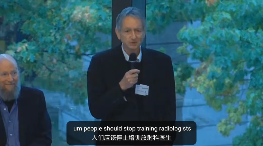
Machine Learning and the Market for Intelligence was organized by the Rotman School of Management at the University of Toronto, Canada.
A decade ago, the scenario Hinton predicted did not occur. The shortage of radiologists is still a real problem facing the medical system. Although AI has achieved expertise in many imaging tasks, it is still difficult, like doctors, to comprehensively examine multiple organs to identify a variety of concurrent abnormalities.
Now, the Aridamo Institute has given a new answer to this question.
On September 18, DAMO RADAR, a general medical imaging model developed by institutions such as the First Hospital of the Unified University of Zhejiang Medical School in Aridamo, was officially released as an open source scientific paper.
This is also a rare case of Chinese AI models appearing on the cover of Science magazine in recent years.
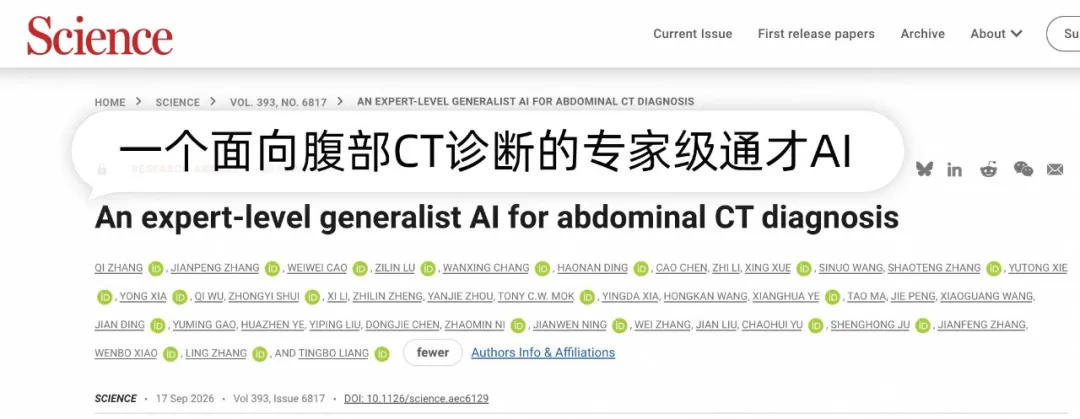
The paper is linked to: www.science.org/doi/10.1126/science.aec6129
This is an open-source address:
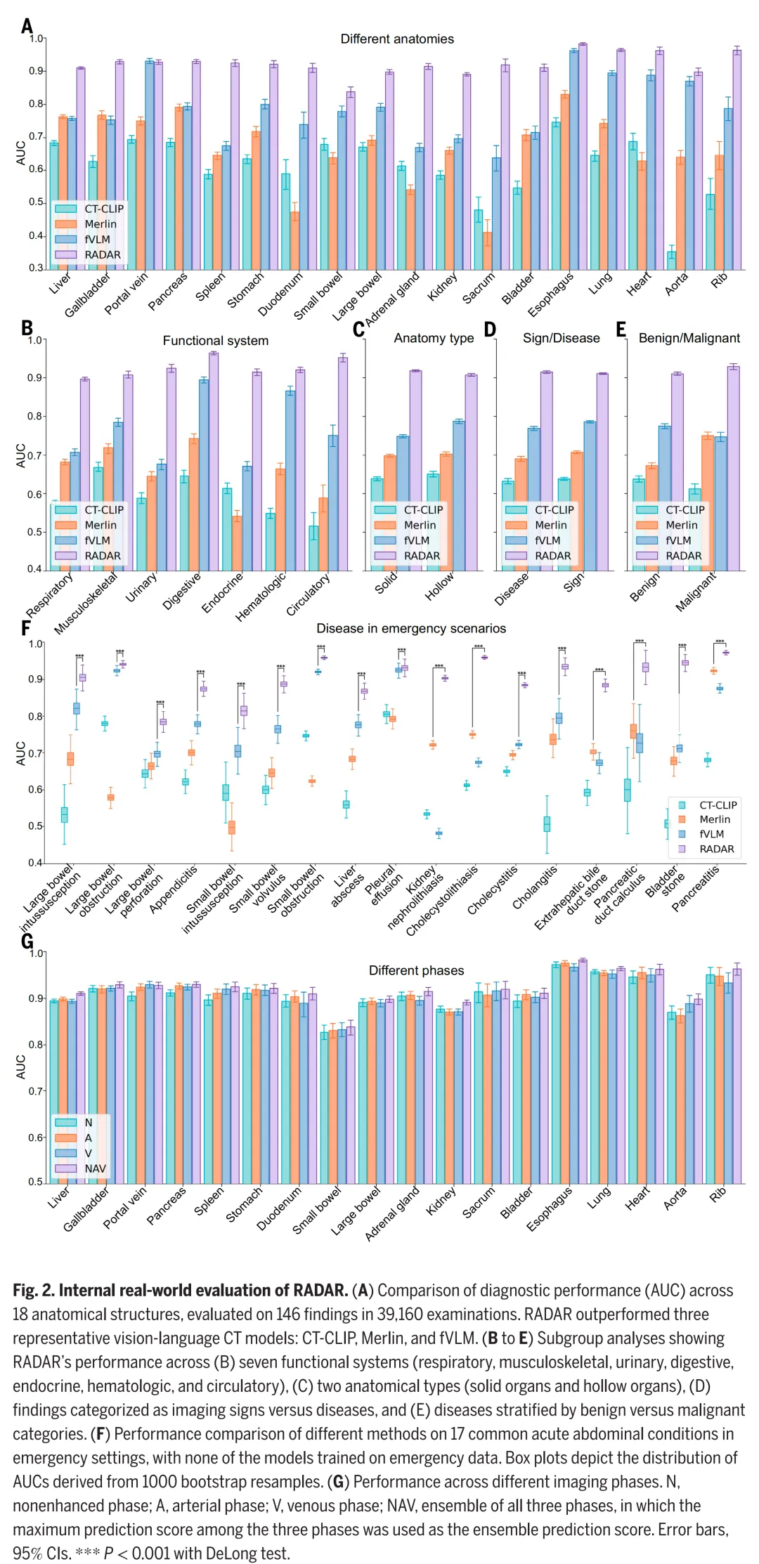
RADAR, also known as Rapid Abdominal Diagnosis with AI and Radiology, is a rapid abdominal diagnosis based on AI and radiology in Chinese. The model focuses on abdominal enhanced CT, and in nearly 40,000 systematic assessments covering 18 anatomical structures and 146 clinically related imaging performances, the average AUC (ability indicator for distinguishing patients from non-patients, 1 for perfect differentiation) is 0.913, the first to reach the level of an imaging expert.
It is even more remarkable that this breakthrough at Damo is not a medical test result from the General Chat Model, which faces serious clinical challenges, with tens of thousands of cases in multiple hospitals and large-scale physician-controlled readings.
Does this mean that medical imaging AI is close to its own GPT moment?
The GPT moment in medical imaging is the moment of the GPT moment, the moment of the GPT moment in medical imaging.
Why are you late?
The medical field continues to attract AI companies. IBM Watson has attempted to provide decision-making support for cancer treatment; DeepMind has explored clinical warning and eye imaging; and in 2026, OpenAI launched ChatGPT Health and OpenAI for Healthcare.
These explorations target different aspects, but face similar requirements. Medical AI must be accurate, reliable, and capable of being reviewed. A missed diagnosis may delay treatment, and a misreport may trigger additional checks. After a high score on a test set, the model still needs to prove its ability to adapt to different hospitals, equipment, scanning protocols, and patient populations.
Image AI is one of the most mature and fast-paced directions of development. In the past, deep learning has driven a number of specialized models that have reached even higher levels of expertise in tasks such as pulmonary node, diabetic retinopathy, and specific cancer screening. These results largely revolve around specific diseases and tasks, collecting images, producing labels, and training models to determine the existence of abnormalities.
When it comes to real clinical tasks, the mission boundaries are often more complex. High scores in single tasks are difficult to directly convert into comprehensive diagnostic capabilities. Diseases covered by models are limited by training labels. With each additional task, data reorganization, labeling, training and verification may be required. The image collection and patient distribution of different hospitals varies, changing an environment, and model performance may decline.
The first single-disease AI project in which senior algorithm expert Zhang Jianqiang at Damoh University participated was lung cancer, followed by a succession of specialized disease projects such as bowel cancer. According to the traditional process, a disease release from position to capacity often takes two to three years. By the time of the second project, the team had found it difficult to cover the real clinical pathway. "Thousands of diseases in the real world, at this rate, may not be complete all the time".
Dr. Zhang, a senior algorithm expert at the Damota Institute, mentions another disadvantage. When the team was developing a pancreatic cancer screening model, it found that AI was able to identify subtle tumour clues, but could still be interfered with by abdominal fluid, post-operative changes or pseudo-positives, resulting in false positives. Abnormal changes that were not usually difficult for doctors to identify, but could instead become blind spots for specialized models. In clinics, it was difficult for the team to develop a model for each interfering factor. The real world is not an AI test, and patients are not likely to have a specific disease, but rather an uncertain disease, a problem that multiple specialized models cannot be solved together.
These difficulties have prompted DAMO RADAR to rethink its approach to learning. The DAMO RADAR, released today, answers a larger question: Can the same model study the relationship between different anatomical structures, imaging representations, and clinical descriptions to cover a broader spectrum of diseases?
Vision-Language Learning provides a new gateway. Clinically produced CT imaging and radiology report natural correlations, and models can directly study these correlations, reducing the need to specify labels for each disease on a case-by-case basis. However, it is still not enough to simply match the entire set of CT and the entire report.
CT imaging and reporting
In terms of anatomical structure, alignment
In order to learn the language of medical imaging, AI systems must first solve a problem: how to accurately match the structure, abnormalities and descriptions in the image with the report? DAMO RADAR-facing abdominal enhanced CT shows the difficulty of this task.
Dr. Shwenpo, director of the Department of Radiology at the University of Tokyo, said that a clinical abdominal enhanced CT usually contains multiple phases, hundreds of images. The abdomen is involved in multiple systems such as digestion, urine, reproduction, and many organs are densely close to soft tissue, and the intestine is surrounded by them. Some diseases differ very little from the surrounding tissues, and doctors need to look at them in stages, combining position, shape, and enhanced performance to make a diagnosis.
For the model, there is another difficulty. Local pathogens in a CT set may represent only a small portion of medical 3D imaging. The report describes multiple organs, both normally seen and abnormal. Directly aligns the entire CT set with the entire report, and critical abnormalities are easily diluted by unrelated information. The model may not be able to accurately match an organ with its abnormalities even after learning the overall association.
DAMO RADAR refined the basic units of learning into anatomical structures. The team matched CT body data with corresponding radiological reports to form a million-level individual data image matching text. Then, using the Qwen language model, the reports were broken down into descriptions of the corresponding anatomical structures and matched with corresponding image regions to obtain more than ten million-level anatomical image matching text.
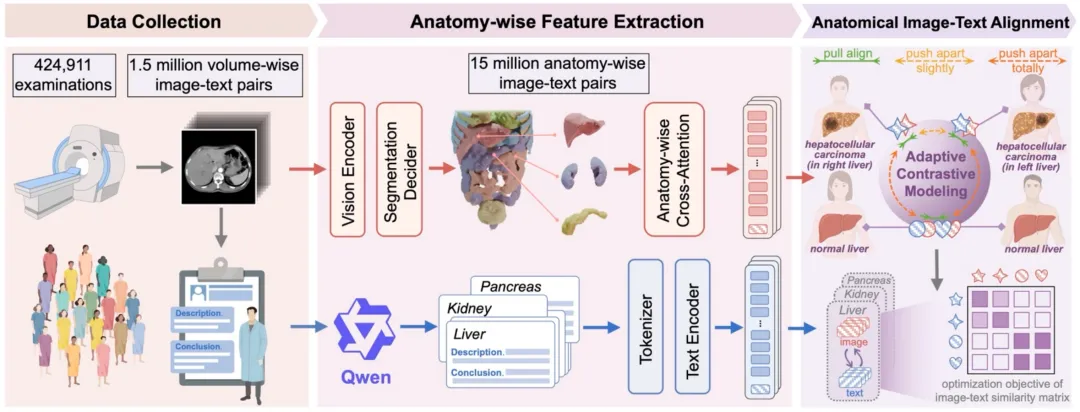
From data collection to extraction of features based on anatomical structures to anatomical-level images - text alignment.
In most cases, clinical reports have been found to provide rich learning signals to reduce the need for disease-by-disease artificial labeling. However, there is a need to distinguish between disease descriptions and monitoring information for the use of anatomical positioning. Models are still divided into pre-trainings on TotalSegmentator datasets with anatomical-grade labeling, resulting in 18 target anatomical structures. At the same time, publicly available TotalSegmentator models are used to generate organ masks as follow-up labels.
The first key link is the alignment of anatomical perception with fine-grained graphs.
As shown below, DAMO RADAR consists of the visual branch and the text branch. The visual branch uses the three-dimensional U-Net to extract image characteristics, and the split decoder is responsible for locating the different anatomical structures. The model aggregates the visual characteristics of the corresponding regions according to the results of the split. The text branch uses the BERT encoder to process the corresponding descriptions in the report. The two types of characteristics are aligned in the same representation space, allowing the model to learn the connection between the visual performance of specific structures and the clinical language.
Zhang Jiansheng mentioned that this way of organizing is closer to the practice of radiation surgeons. They usually examine the liver, pancreas, bile duct, kidney and intestines in sequential anatomical structures and then aggregate information from different parts to make judgments. For DAMO RADAR, clear local responses help to reduce interference with other organs and unrelated descriptions.
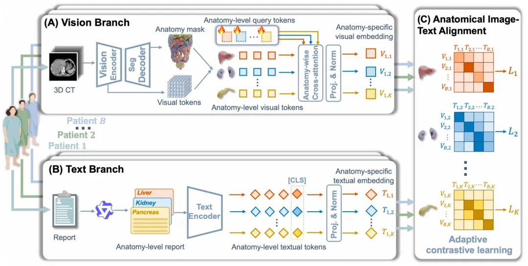
DAMO RADAR architecture overview
The second key link is adaptation versus modeling. After completing local mating, the model also deals with similarities between different patients.
Compared to standard graphing studies, typically paired images are drawn closer to text, and other non-paired samples are considered negative samples. However, different patients do not necessarily represent different imaging meanings. The normal liver of two patients may have similar characteristics, and similar abnormalities described may also share some imaging performance.
DAMO RADAR, based on the normal or abnormal state of the anatomical structure, and the semantic similarity between abnormal descriptions, dynamically adjusts the supervisory relationship of learning versus learning. For normal structures, samples between different patients can serve as additional samples. For abnormal structures, the similarity of models based on text descriptions, by gently supervising the adjustment of mating weights, reduces the cases where a semantic sample is over-propelled.
After the training is completed, the model uses text prompts to judge the performance of the target image. For example, colonoscopy, the input describes the presence and absence of the prompts, respectively, and the similarity with the imaging characteristics of the corresponding anatomical structure is predicted. The disease query can therefore be replicated using the same set of diagrams, without having to re-train a special classifier for each query.
The anatomical level is aligned to solve the problem of the scarcity of diagnostic signals in 3D imaging, while adapting to the disruption caused by learning to further reduce semantic similarities between patients. The combination of the two enables the same model to cover a wider range of imaging performance and to query different objectives through text suggestions, reducing the cost of disease-by-disease labeling and repeat training. For the entire industry, long-term accumulated imaging and reporting data from hospitals have the opportunity to transform into continuously expanded diagnostic capabilities.
It's a good idea to get out of the training data.
Multi-Centered and Real Reading Screening
CT imaging and reporting align at the anatomical structural level, solving the problem of model learning. Two more practical questions follow: Can models maintain performance in untrained hospitals?
Look at the internal real-world tests. The team used a sequence of cases over an independent period of time to construct a test queue that included 39,160 abdominal enhanced CT scans, covering 18 anatomical structures and 146 clinically related imaging performances. The results showed that the average AUC of DAMO RADAR was 0.913.95%, with confidence ranging from 0.911 to 0.915.
Classification results showed that DAMO RADAR performed relatively well on different missions. The AUC for 18 anatomical structures ranged from 0.838 to 0.982.[4] The average AUC for substantial organs and cavity organs was 0.918 and 0.907, respectively, and the average AUC for disease and imaging symptoms was 0.914 and 0.911 respectively.[5] The average AUC for good and bad performance was 0.910 and 0.929 respectively.[6]
These results show that the model's capabilities are not concentrated in a few high-frequency diseases or certain types of organs, but that it is able to process substantial organs such as the liver, pancreas and to identify abnormalities in cavity organs such as the gastrointestinal tract at the same time.
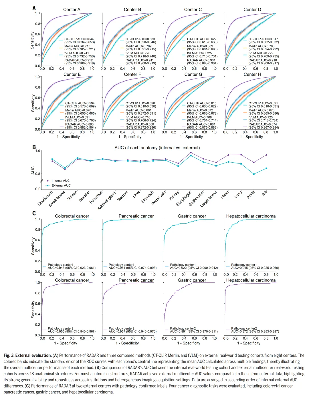
One of the most challenging aspects of the model was whether it could remain stable when faced with different hospital equipment, scanning programs, and patient composition. The team tested at eight external centers, including 24,239 enhanced abdominal CT scans. These cases came from different scenarios such as emergency rooms, hospitals, outpatient clinics, and health clinics, with scans covering the entire abdomen, upper abdomen, lower abdomen, and pelvic cavity. The number of imaging performances evaluated by each center ranged from 68 to 136.
The results are still prominent, with DAMO RADAR having an average AUC of 0.895 at eight external centers, significantly superior to the second-ranked fVLM, and a single-centre AUC of between 0.874 and 0.912. The average AUC decreased by 0.018 compared to the internal queue. Performance decreased, but the centers remained relatively stable. More than half of the anatomical structures in internal and external tests differed by less than 0.01 in AUC, including areas such as the stomach, liver and esophagus.
The team also set up several groups of higher difficulty tests.
The training goal excluded the emergency room, and the team used 27,267 emergency room examinations to evaluate DAMO RADAR's ability to identify 17 common acute abdominal disorders. The average AUC of the model was 0.904.95% in the confidence range of 0.900 to 0.909.
Cross-crowd verification uses the Merlin test set, which is open to the public at Stanford Hospital. The data set contains 5,137 CT scans, mostly for Western patients. DAMO RADAR does not use Merlin data to train or fine-tune, directly testing 21 imaging performances within its existing capabilities, with an AUC of 0.883.
Another test introduced more stringent pathological diagnostic criteria. Two independent external sequences included a total of 4,333 cases, including 648 colorectal cancers, 388 pancreatic cancers, 537 stomach cancers, 383 liver cell cancers, and 2,377 normal controls. DAMO RADAR's AUC for all four cancers ranged from 0.891 to 0.984.[3] Models were trained primarily through imaging and radiological reports, but maintained high performance in pathologically confirmed cases, providing further evidence for the reliability of their diagnostic signals.
The study included 26 radiologists from 14 institutions and 300 abdominal enhanced CT scans covering 61 types of imaging performances. Doctors were divided into four groups based on their experience and institutions, including senior and junior radiologists from top hospitals and other hospitals.
In the first phase, doctors independently read CT and write reports. The results show that most doctors' workpoints are below the curve of the subjects' work characteristics of DAMO RADAR, with only three senior doctors slightly higher than the model (see Figure A below). However, the comparison here is a mere image reading performance, where doctors do not combine complete medical history, laboratory examinations and other clinical information.
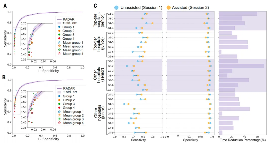
After a wash-out period of at least a month, the same group of doctors re-read the patient in the order of the repeated cases. This time, DAMO RADAR gives predictions and detects the suspect area by attention-graphing. AI-assisted increases the overall sensitivity of the doctor by 10.0%, the specificity slightly decreases from 98.8% to 98.2%, and the average reading time per case decreases by 30.7% (as shown in Figures B and C above). This indicates that the model improves the detection of abnormalities, and the cost of the specificity decreases slightly.
Level results further show that DAMO RADAR helps doctors of different experience levels. In top hospitals, AI-assisted primary care physicians perform similarly to senior doctors who do not use AI. Other hospitals also observed the same trend. AI-assisted primary care physicians narrow the reading gap between primary care physicians and senior physicians.
For general medical imaging AI, these results show that a model is beginning to endure multiple tests at the same time as disease coverage, cross-central generalization, and human-computer collaboration. Of course, current clinical verification is still based on retrospective research. Whether DAMO RADAR can maintain performance in forward-looking clinical environments needs to continue to be verified in more diverse populations and medical settings.
It's all about intelligence.
Breaking the boundaries of "one disease one"
Understanding the significance of DAMO RADAR also needs to be traced back to DAMO's research into medical AI over the past few years. DAMO will focus on "flat-scanning CT+AI", hoping to take advantage of the large number of conventional CT scans available in clinical and medical examinations to detect easily overlooked disease clues.
This pathway was first developed in a specialized disease model. In 2023, DAMO PANDA used flat CT screening for pancreatic cancer, and in a real-world retrospective study of more than 20,000 patients found multiple cases of previously undiagnosed lesions, which was described by the top medical journal Nature & Medicine as "the Golden Age of imaging AI-based cancer screening". DAMO GRAPE will then explore expanding into stomach cancer, and DAMO COCA will further explore the possibility of colorectal cancer screening. DAMO iAorta enters the emergency room, uses flat CT to identify acute coronary syndrome, and receives certification in real hospital workflows.
These projects, covering substantial organs, cavities and acute atherosclerosis, have gradually revealed expanding costs in the process of validating the clinical value of the pathology pathway. DAMO RADAR has completed a key shift: it has begun to use large-scale CT imaging and radiological reporting learning, and no longer relies on the individual production of training labels for each target disease.
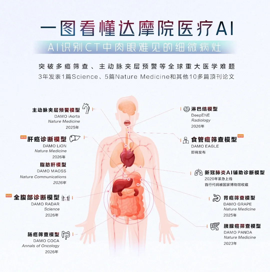
DAMO RADAR has expanded the scope of its research to more complete abdominal imaging. The two types of future models may coexist in the long term. DAMO models are suitable for deep optimization around high-risk diseases, and general models take on a wide range of tasks such as finding abnormalities, assisted reading, and second opinions.
Ten years ago, Hinton envisioned AI replacing radiation surgeons. DAMO RADAR's answer was closer to reality: AI becomes an auxiliary tool for doctors to help them detect more abnormalities and complete the scan in less time.
While the GPT time for medical imaging AI cannot be judged, DAMO RADAR at least clarifies the picture. Its open source provides the conditions for more institutions to reproduce, validate and expand this route.