“ A robot performs fetal surgery, and it runs on sheep.
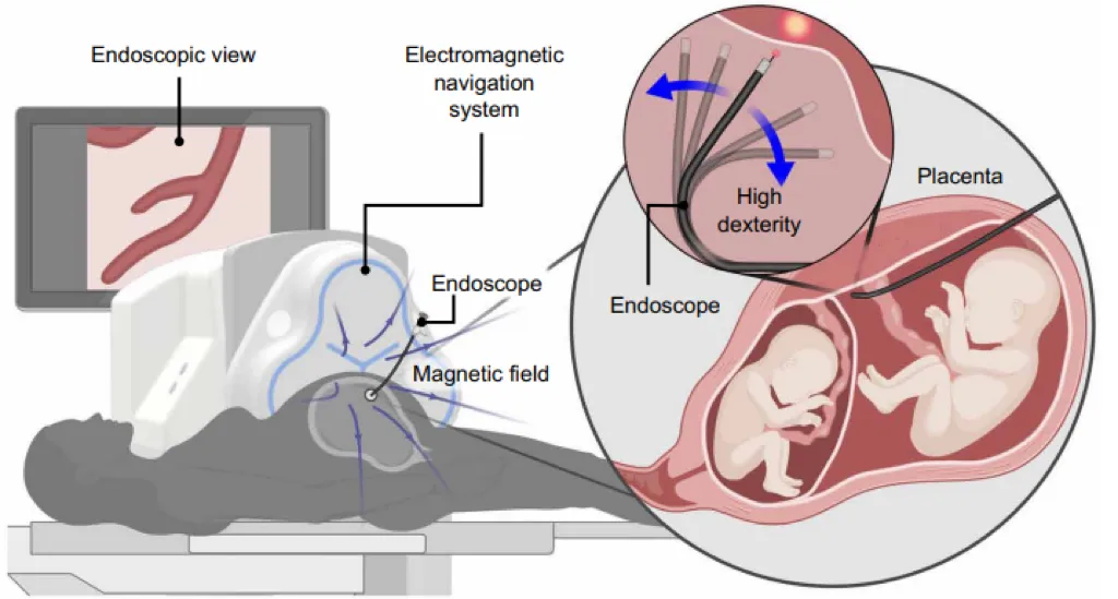
A team of researchers from institutions such as ETH Zurich has just published a new paper in the international peer-reviewed journal Science Robotics: They have developed a magnetically-controlled flexible lens robotic platform and, for the first time, have fully verified its ability to navigate and dissolve in TTTS laser surgery on live sheep models.
The study is worth mentioning because it was the result of a microsurgery of a hard bone with a large open cavity.
01.
Why is this surgery so difficult?
Transfusion syndrome is a deadly disease that affects 10 to 15 percent of pregnancies of twins with a single placenta. The current gold standard treatment is called fetal lens laser gel sterilization (FLC): doctors insert the fetal lens into the uterus through a tube, scan the surface of the placenta to find a matching pair of blood vessels that connect the two fetal blood cycles, and then use a laser to burst them.
The difficulty is that even experienced senior doctors who operate knives in large centers have a survival rate of about 70 percent and about 10 percent of surviving children experience long-term neurodevelopmental damage.
The problem is mainly instrumental. Now clinically used is Karl Storz's pre-curved rigidity foetal mirror (11508AAK), which is particularly sensitive when encountering a large plaque in the front of the uterus (based on the mother's abdomen). The field of view can only be tilted to the surface of the plaque, and the blood vessels are neither visible nor easily burned.
What is even more troubling is that "large open cavities" such as the uterus, stomach and bladder do not have the same tubular walls as the intestine to constrain and stabilize instruments, and flexible instruments are more difficult to control; the vision of the retina is especially small, and doctors can only use the brain to record the entire placenta's blood vessels, which is a huge mental burden.
02.
A 3.2-millimeter "magnetic controlled soft tube"
The team's solution was a robotic foetal lens with a diameter of only 3.2 mm, thin enough to pass through a commonly used clinical 10-Fr tube (which is important, the thicker the tube, the worse the clinical prognosis).
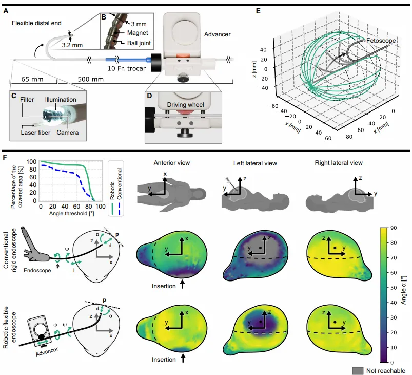
Its flexible end is composed of a series of magnets and precision steel ball joints, which are driven by a magnetic field generated by an external electromagnetic navigation system (eMNS), while the insert depth is controlled by a mechanical propulsion unit.
As a result, the soft lens has a much better range and melting angle than conventional rigid foetal lenses. Doctors consider 90° to be the ideal melting angle, whereas robotic foetal lenses can reach more than 45° in 91% of the frontal position, compared to 73% in conventional instruments; when required above 70°, the coverage of both is 89% and 33%, respectively, and the gap is very large.
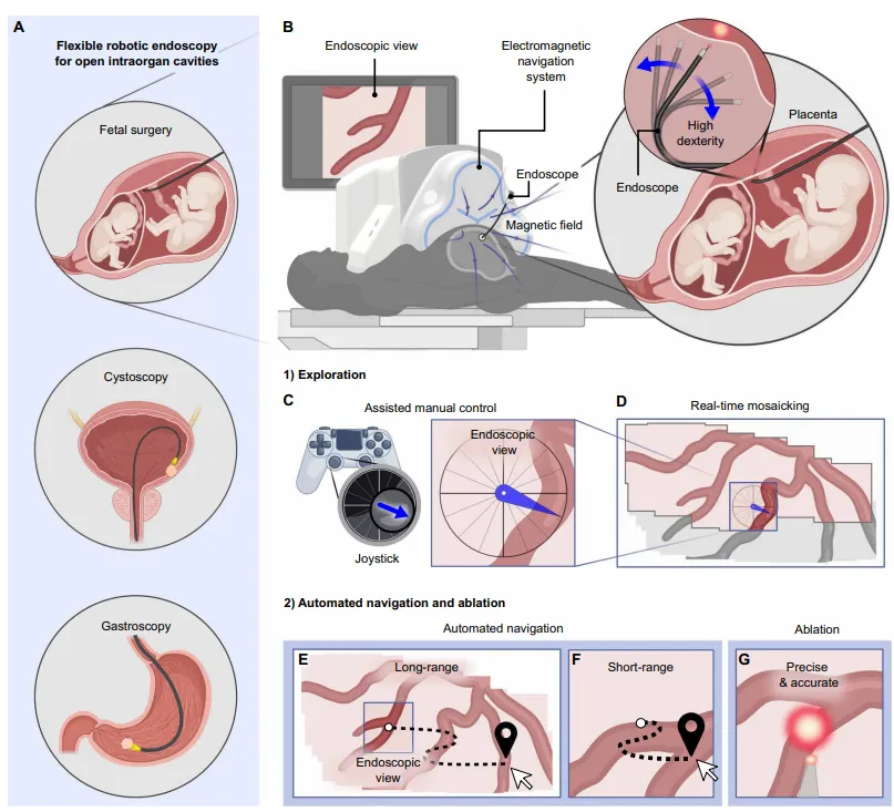
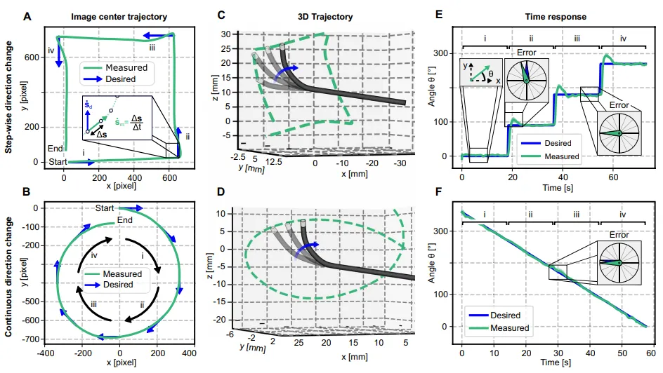
The control method is another highlight of the system: it is entirely image-based. It relies on visual servers to move the desired direction of the target point in the interior lens image, mapping to the magnetic field, without the need for additional positioning sensors to be embedded in small lenses. The method is "modelless" and does not rely on complex robotic modeling, so it can be adapted to devices of varying soft hardness.
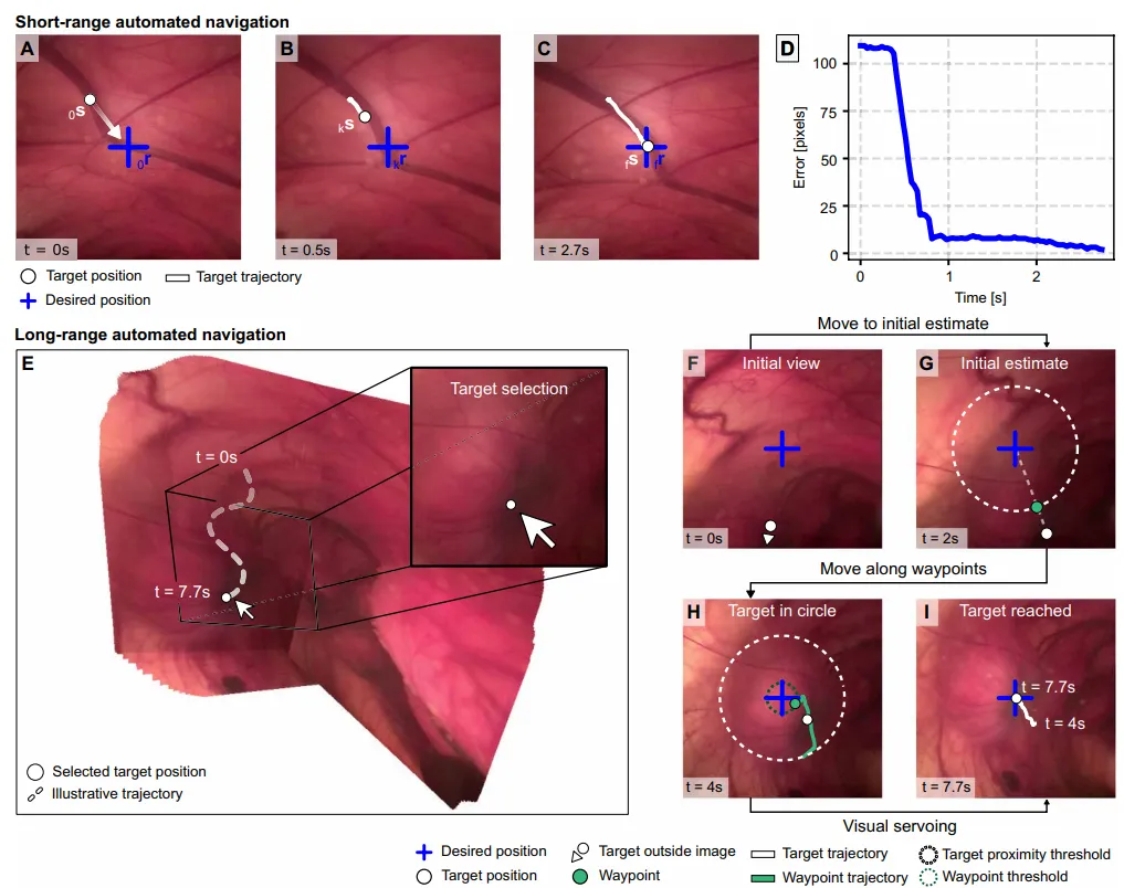
The team devised three navigation strategies to match the different stages of the surgery: manual navigation of the moving image directly controlled by the doctor with a handshake; automatic short-range automatic navigation of the targets in the field of view to the center of the image; and selection of targets outside the current field of view on the image collage map that has been generated; and automatic navigation of past long-range automatic navigation.
03.
Running the whole process on a live sheep
The team used a pregnant black goat (about 50 kg, 90 to 100 days pregnant) to make a model of a female sheep that had similarities to humans in placenta and newborn development, the closest to the real model before clinical transformation.
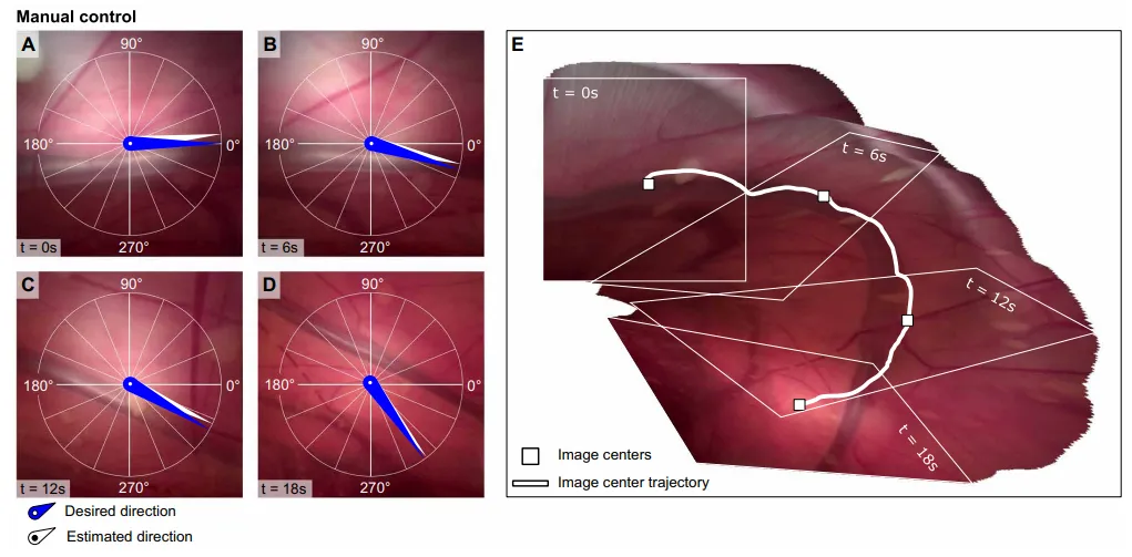
An experienced fetal surgeon used a rocking rod to navigate the vessels along the uterine wall, with an average of 8.6 degrees of angular error between the expected direction and the actual estimated direction, and the system also collated the accompanying image into a real-time panorama. In short-range automated navigation, all six targets were steadily sent to the two-pixel error at the center of the image, taking an average of about 3.1 seconds; long-range navigation had an accurate impact on the selected visual field targets in the collation chart, reaching an average of 6.31 seconds, and reaching a distance from the starting point to the target.
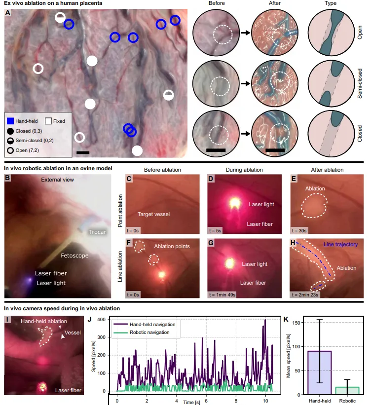
The decomposition link also illustrates the value of the robot. The team first made controls on the dissociated human placenta: under the same laser power and duration, traditional foetal mirror replicas in hand could not completely burn one of the seven blood vessels, while the method of fixing the laser fiber ("simulating the robot to remain stationary") in seven of them burned three, half-closed two.
In the live-sheep experiment, doctors completed point melting and line melting (Solomon's technique), which simulates the connections that have coalesced into branches. Thanks to the filter film on the camera, blood vessels are still clearly visible when the laser is working, avoiding sensor over-exposure, which is precisely what previous similar studies have failed to reliably show melting card dots. Compared to handheld and robotic methods, the robot navigates the image at a speed of 16 pixels per second, while with up to 90 pixels per second, the laser burning traces are more concentrated.
04.
In addition to fetal surgery, beginners are also good.
To verify the ease of use, the team also recruited 11 participants (one of whom was a fetal surgeon with 25 years of experience in a lens, the rest being beginners) for control tests.
The results showed that in the simulation of the melting point of a single matching joint, the melting accuracy median of the robotic embryos was 140 micrometers, compared to 621 micrometers for conventional instruments; in the 302 micrometers versus 451 micrometers, the accuracy and stability were significantly better. The cost was that the robot moved slower between targets (approximately 20 seconds vs 8.5 seconds).
At the same time, the navigation and splicing method is not limited to fetal surgery. The team uses magnetic control lenses of different sizes, different softness, and on the isolated pig bladder and exoskeleton stomach models, also verified the manual operation of the bladder and stomach lenses, automatic navigation with real-time splicing, and even with live screening samples.
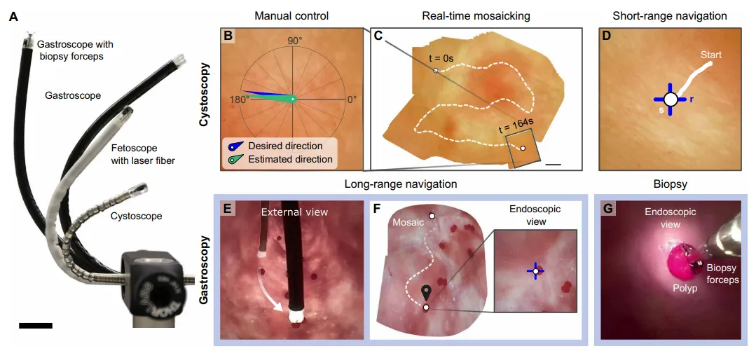
Of course, the team also pointed out the limitations: in physical experiments, splicing is limited in size, long navigation distances are limited; in the future, it will be necessary to improve the robustness of splicing algorithms against shadows (such as moving fetuses) and add reliable machine positioning and real-time 3D reconstruction.
Overall, this work, which combines magnetic-controlled flexible robots, image navigation and real-time landscape pairing, completes a core process of complex fetal surgery in a live large mammalian model and is a solid step towards the clinical transformation of TTTS robot therapy.
Address of the paper: