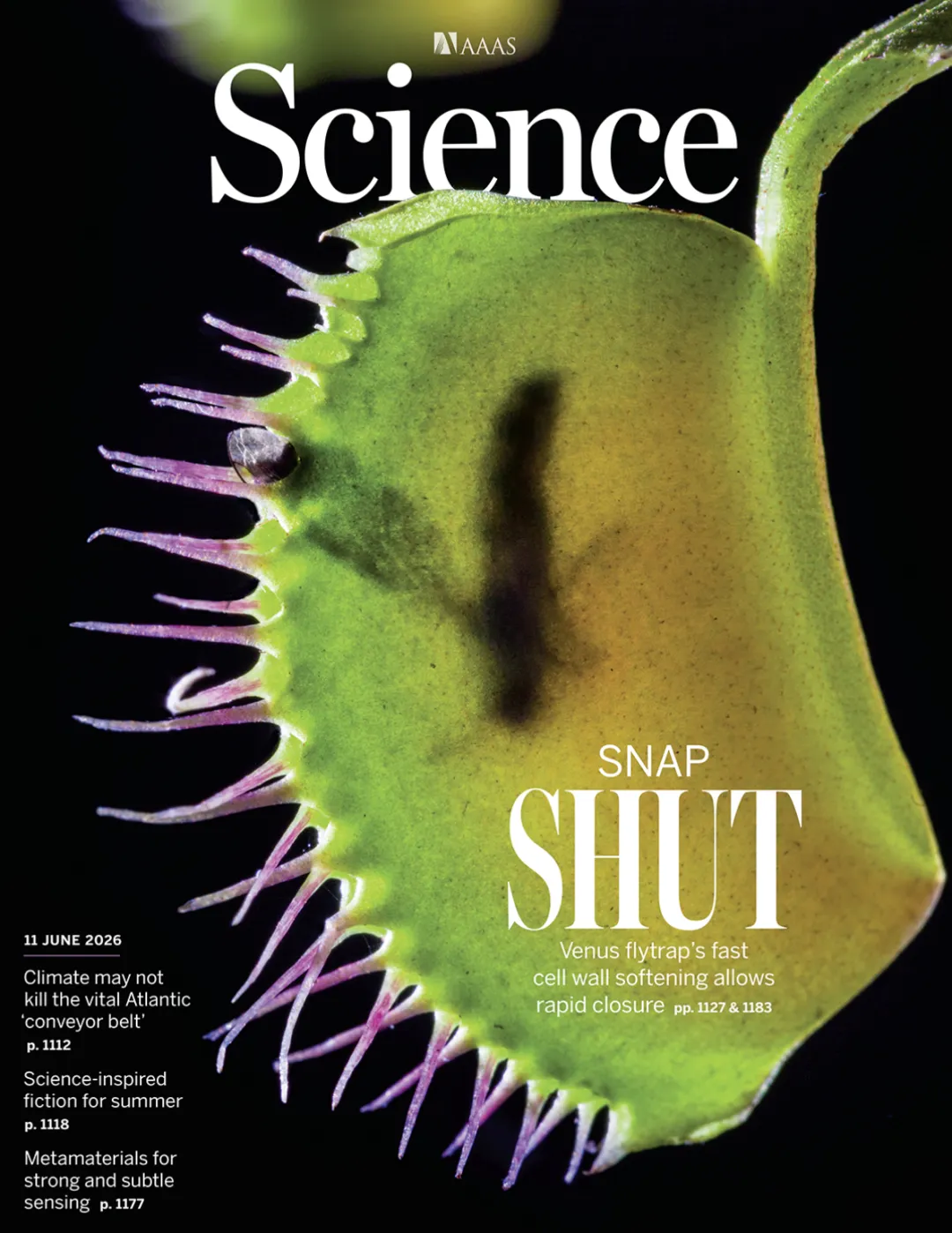
“ With no muscles, no gills, and only 0.1 seconds to catch the herring, the insect catcher relies on turning its local gills into soft cuts, a type of drive that software robots have not yet used.
The outer layer of plant cells is covered by a hard, heavy cell wall of cellulose. This means that the animal's plan to change the shape of the cell based on myoproteins - myoproteins - is basically not working in plants.
Its caterpillars consist of two leaflets and a central artery in the middle, and when the surface is touched twice in a short period of time, the action electrodes propagate along the leaf, followed by about 0.1 seconds, and the caterpillars merge.
This phenomenon has been studied for more than a century since the time of Darwin. The long-standing mainstream explanation is "water": penetration drives water to flow in the direction of the thickness of the leaf, resulting in different degrees of inner and outer surface expansion.
A team of researchers from France ruled out this explanation with a set of prototype measurements. They have been measuring subcellular scales from whole organs, using single-cell pressure probes, tissue swelling experiments, prototype nanoscapes and diffuse co-focusing contourizers, and concluded on the cover of the SiliconScience paper: The real drive for the closure of insect larvae is the rapid softening of the outer skin cell walls at about a second's scale. This is believed to be the fastest mechanical change currently reported in plant cell walls.

01.
0.1 seconds, which is not really the speed of Madha.
To find the source of the drive, the first step is to figure out who exactly owns the 0.1 second figure.
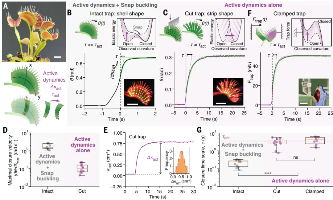
The team first used a 3D visual device to track the entire closing process of the insect prey. The curve shows that the closing initially underwent a slower acceleration, followed by a rapid turn of the leaf curve from convex to convex, with a characteristic time of about 0.21 seconds and a maximum angular velocity of about 1.88 rad/s.
This is typical of snap-buckling. Insect-hunting is a double-curved, elongated structure that bends and stretches together to form an energy barrier. Plants need to slowly penetrate the bulb into the tissue, and once it crosses the threshold, the entire structure passively releases energy at once. In other words, the speed is magnified by the geometrical structure, and in 0.1 seconds a considerable part of the function belongs to the structure itself, not to the internal drive unit.
How fast was the drive unit itself? The team used two complementary routes.
The first penetration: cut the blade vertically to the midbrain. Thus the curve and stretch are dissolved, the impact barrier is removed, and the remaining movement directly reflects the change in the internal active curvature. The cut itself will immediately trigger closure, but after the insect larvae are restored, they can be reopened and stimulated again. The maximum angle measured is about 0.12 rad/s, one degree slower than the full larvae, and the change in the active curvature is about 0.5 cm per second, measured in about 3.1 seconds.
The second is unbreakable: the full state of the open insect prey is placed between two rigid walls, one of which is fitted with a force sensor to record the force required to maintain the fixed distance between the leaves of the prey. The shape of the prey prey is locked, the stretch remains constant, and the change in force is directly proportional to the dynamics of the active curvature. The measured time scale is about 4.1 seconds, consistent with the cutting experiment.
Both approaches give the same answer: the internal drive of the grass catcher is measured in 3 to 4 seconds, and then amplified by the unstable structure of the grass to become the "grass" seen with the naked eye.
This has reference value for driver design: fast action can be achieved by a combination of "slow drive unit + programmable unstable structure", and the drive itself does not have to respond in milliseconds.
02.
The speed of the water is almost one degree.
With "a few seconds", the standard hydromechanical hypothesis can be quantified.
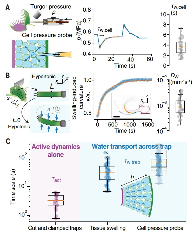
The team first used a single-cell pressure probe to directly measure the water exchange process of large thin-tissue cells inside insect larvae. The result was a single-cell water transfer time of about 3.7 seconds, and the corresponding membrane permeability was close to the reported upper limit of plant cells.
The problem arises in the length of the pathway. Water must pass through the entire thickness of the leaf, with about 5 to 10 layers of thin-walled cells separated in the middle.
In order to cross-validate, the team performed a penetration melting experiment on the organization scale. They first identified a one-to-one correspondence between the curvature of the tissue layer and the water pressure, i.e. the degree of curvature can act as an agent indicator of the amount of water absorbed; then quickly transferred the tissue layer from 0.4 M of the citrus alcohol solution to 0.1 M of the solution, and the curvature dynamics of the melting process developed an effective water dispersion coefficient.
The two sets of completely independent measurements point to the same conclusion: the transmission of water is slower than the active curve by almost a quantitative level.
There is also a geometric proof. The time of diffusion is proportional to the square of the thickness, and the thickness of the insect prey from the edge to the median is gradual. If the closure is driven by water transmission, the curve change should show a space delay in propagation from the edge to the median. This delay is not observed in experiments.
Long-distance water transport is therefore excluded from the main driving mechanism.
03.
The real driver is a layer of skin cell wall.
The remaining candidate is the mechanical nature of the cell wall itself. This assumption was made decades ago, but has been lacking in direct evidence.
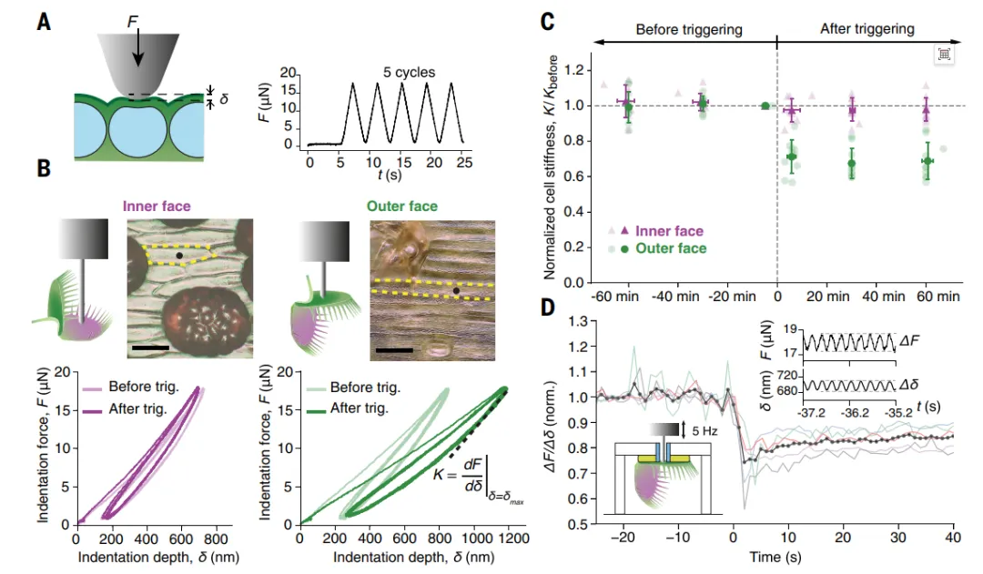
The team used zinc to attach the still-active insect prey to an open state, triggering the same batch of epithelial cells by matching the original nanoscale scratches, thus excluding the interference of geometrical changes.
The data differentiation is high: the inner surface skin hardness is almost unchanged, the outer skin hardness significantly decreases, after triggering it decreases to about 0.69 times the original, a decrease of about 30%, and this softening lasts at least an hour.
The two controls are equally critical. When switching to a millimetre-level probe (which detects the depth of internal tissue), the rigidity of both sides does not change significantly, indicating that the softening is limited to this layer of the outer skin; and softening is not enough to trigger movement in the event of just one touch, which means that a single action current or single wave of filtration is not enough to cause this mechanical change.
However, the above measurements were carried out within minutes of triggering, and there was no causal relationship. So the team set up a pressurizing device that could track the stiffness in real time: restraining the motion mechanics of the insect prey within a few microns, overlapping the high frequency force at a constant load. The results of the measurements were that the stiffness of the outer skin cells began to decrease within a few seconds of triggering, and the time scale matched the active curvature time specified on the front mark.
The final difficulty was to distinguish between "softening of the cell wall" and "softening of the pressure drop". Both of these things were difficult to distinguish in the scratch data. The team changed their minds and took advantage of the differences in the direction of the shape response: for a cell that is full of expansion, a decrease in the pressure drop will make the outer wall beat less, while the softening of the cell wall will make it beat more. This judgment is determined by force balancing, which has been tested experimentally and numerically.
They used dynamic composite mixed-color scatter-focused rotor meters to measure the surface shape of the outer skin at a sub-micrometer high resolution. When triggered, the surface "inflammation" increased systematically by about 8%, from about 595 nanometers to about 647 nanometers, the equivalent of several tens of nanometers.
Putting cell geometry, inflation data and compression curves together into a finite metamorphic modeling inversion, a set of specific parameters is obtained: inflation of about 0.77 MPa, a decrease of about 40 percent from 64 MPa to 37 MPa. Further consideration of tissue stress and cell wall heterogeneity, the decrease is about 30 percent, and the trend is consistent.
In the open state, the bulging of the epithelial cells stretches both inner and outer layers of the epithelial skin, with a pre-stress of about 0.06 (pushed by mass-wall separation experiments); after a decrease in outer wall modulus, the epithelial skin is partially expanded relative to the inner surface of the epithelial skin, resulting in an active curvature of about 0.5 cm per inch and an increase in the number of test points. The curvature occurs mainly in the direction of the vertical medial artery, because the epithelial cells have a strong expansion in the circumference, so that the pressure of the epithelial skin is inversely proportional.
So far, the entire chain has been closed: the outer skin cell wall is rapidly softened, releasing the elastic predisposition stored in the enlarged epithelial cells, driving the activated curvature of the epithelium, and being accelerated by the unstable structure of the epithelium.
For the field of robotics, this work gives a driving pattern that has not been clearly identified before: by localizing material rigidity, releasing elastic stresses that have been previously stored at equilibrium. It bypasses the physical limitations of fluid transport speeds and improves the difficulty of controlling pure mechanical instability. The paper clearly lists these two as the advantages of the mechanism over existing solutions, and points to non-muscle fast drivers, software robots and stimulus response materials.
To be more precise, the way to drive the work can be done in addition to the transfer of fluids, the transformation of the volume, and the transformation of the soft hardness of the material. The design of the transformed rigidity structure and the pre-stress storage of energy is an idea that can be directly borrowed.
Address of the paper: